- Volumes 84-95 (2024)
-
Volumes 72-83 (2023)
-
Volume 83
Pages 1-258 (December 2023)
-
Volume 82
Pages 1-204 (November 2023)
-
Volume 81
Pages 1-188 (October 2023)
-
Volume 80
Pages 1-202 (September 2023)
-
Volume 79
Pages 1-172 (August 2023)
-
Volume 78
Pages 1-146 (July 2023)
-
Volume 77
Pages 1-152 (June 2023)
-
Volume 76
Pages 1-176 (May 2023)
-
Volume 75
Pages 1-228 (April 2023)
-
Volume 74
Pages 1-200 (March 2023)
-
Volume 73
Pages 1-138 (February 2023)
-
Volume 72
Pages 1-144 (January 2023)
-
Volume 83
-
Volumes 60-71 (2022)
-
Volume 71
Pages 1-108 (December 2022)
-
Volume 70
Pages 1-106 (November 2022)
-
Volume 69
Pages 1-122 (October 2022)
-
Volume 68
Pages 1-124 (September 2022)
-
Volume 67
Pages 1-102 (August 2022)
-
Volume 66
Pages 1-112 (July 2022)
-
Volume 65
Pages 1-138 (June 2022)
-
Volume 64
Pages 1-186 (May 2022)
-
Volume 63
Pages 1-124 (April 2022)
-
Volume 62
Pages 1-104 (March 2022)
-
Volume 61
Pages 1-120 (February 2022)
-
Volume 60
Pages 1-124 (January 2022)
-
Volume 71
- Volumes 54-59 (2021)
- Volumes 48-53 (2020)
- Volumes 42-47 (2019)
- Volumes 36-41 (2018)
- Volumes 30-35 (2017)
- Volumes 24-29 (2016)
- Volumes 18-23 (2015)
- Volumes 12-17 (2014)
- Volume 11 (2013)
- Volume 10 (2012)
- Volume 9 (2011)
- Volume 8 (2010)
- Volume 7 (2009)
- Volume 6 (2008)
- Volume 5 (2007)
- Volume 4 (2006)
- Volume 3 (2005)
- Volume 2 (2004)
- Volume 1 (2003)
• Gold nanoparticles were successfully synthesized from Plumeria alba flower extract.
• The size of the gold nanoparticles could be controlled by varying concentrations of flower extract.
• Small size gold nanoparticles exhibited better antibacterial and catalytic activity.
• Mechanism of catalysis was explained.
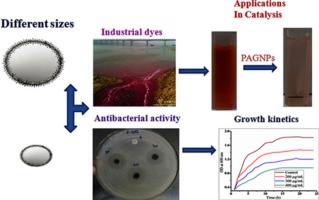
Bio-inspired eco-friendly gold nanoparticles were synthesized by a green method using aqueous Plumeria alba flower extract (PAFE). The use of 1% and 5% concentrations of PAFE resulted in two different sizes of P. alba gold nanoparticles, PAGNPs1 and PAGNPs2, with surface plasmon resonance (SPR) peaks at 552 and 536 nm, respectively. Size-controlled formation of gold nanoparticles was indicated by the SPR shift observed with increasing concentration of PAFE. The accurate size and morphology of PAGNPs1 and PAGNPs2 were determined by transmission electron microscope (TEM) analysis is found to be 28 ± 5.6 and 15.6 ± 3.4 nm, respectively, and those are spherical in shape. The antibacterial activity of PAGNPs1 and PAGNPs2 was tested against Escherichia coli; the small-sized PAGNPs2 exhibited better antibacterial activity with a 16-mm zone of inhibition at a concentration of 400 μg/mL. Furthermore, the catalytic activity of PAGNPs1 and PAGNPs2 was analyzed on six hazardous dyes; PAGNPs2 exhibited more pronounced catalytic activity than PAGNPs1. Among all of the dyes, 4-nitrophenol was most rapidly degraded to 4-aminophenol by PAGNPs2 within 5 min. The mechanism of catalysis in the presence of PAGNPs1 and PAGNPs2 can be described as an electron transfer process from donor NaBH4 to an acceptor. The facile green synthesis of such eco-friendly nanoparticles in bulk suggests this method has potential industrial applications.

