- Volumes 84-95 (2024)
-
Volumes 72-83 (2023)
-
Volume 83
Pages 1-258 (December 2023)
-
Volume 82
Pages 1-204 (November 2023)
-
Volume 81
Pages 1-188 (October 2023)
-
Volume 80
Pages 1-202 (September 2023)
-
Volume 79
Pages 1-172 (August 2023)
-
Volume 78
Pages 1-146 (July 2023)
-
Volume 77
Pages 1-152 (June 2023)
-
Volume 76
Pages 1-176 (May 2023)
-
Volume 75
Pages 1-228 (April 2023)
-
Volume 74
Pages 1-200 (March 2023)
-
Volume 73
Pages 1-138 (February 2023)
-
Volume 72
Pages 1-144 (January 2023)
-
Volume 83
-
Volumes 60-71 (2022)
-
Volume 71
Pages 1-108 (December 2022)
-
Volume 70
Pages 1-106 (November 2022)
-
Volume 69
Pages 1-122 (October 2022)
-
Volume 68
Pages 1-124 (September 2022)
-
Volume 67
Pages 1-102 (August 2022)
-
Volume 66
Pages 1-112 (July 2022)
-
Volume 65
Pages 1-138 (June 2022)
-
Volume 64
Pages 1-186 (May 2022)
-
Volume 63
Pages 1-124 (April 2022)
-
Volume 62
Pages 1-104 (March 2022)
-
Volume 61
Pages 1-120 (February 2022)
-
Volume 60
Pages 1-124 (January 2022)
-
Volume 71
- Volumes 54-59 (2021)
- Volumes 48-53 (2020)
- Volumes 42-47 (2019)
- Volumes 36-41 (2018)
- Volumes 30-35 (2017)
- Volumes 24-29 (2016)
- Volumes 18-23 (2015)
- Volumes 12-17 (2014)
- Volume 11 (2013)
- Volume 10 (2012)
- Volume 9 (2011)
- Volume 8 (2010)
- Volume 7 (2009)
- Volume 6 (2008)
- Volume 5 (2007)
- Volume 4 (2006)
- Volume 3 (2005)
- Volume 2 (2004)
- Volume 1 (2003)
• CdS nanoparticles with a mean particle size below 10 nm were synthesized by microwave irradiation.
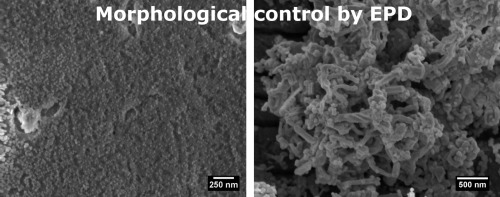
• CdS nanostructures were formed by electrophoretic deposition (EPD).
• Morphology of CdS nanostructures could be controlled by the synthesis and deposition conditions.
• Thin films and one-dimensional nanostructures could be obtained by this methodology.
Thin films and one-dimensional CdS nanostructures were formed by electrophoretic deposition of nanoparticles with morphological control. Particles less than 10 nm in diameter were prepared by a microwave assisted synthesis using trisodium citrate as capping agent. The particles were characterized by UV–Vis spectrophotometry, dynamic light scattering, and X-ray diffraction. Nanoparticles in the resulting aqueous dispersion were electrodeposited without any further treatment on aluminum plates. The nanostructures consisted of thin films with surface roughness being tuned with the applied voltage. One-dimensional nanostructures were formed by varying the applied voltage and by changing the molar ratio of the precursors in the nanocrystal synthesis. These results illustrate two different mechanisms of electrophoretic deposition that lead to the formation of different nanostructures.

