- Volumes 84-95 (2024)
-
Volumes 72-83 (2023)
-
Volume 83
Pages 1-258 (December 2023)
-
Volume 82
Pages 1-204 (November 2023)
-
Volume 81
Pages 1-188 (October 2023)
-
Volume 80
Pages 1-202 (September 2023)
-
Volume 79
Pages 1-172 (August 2023)
-
Volume 78
Pages 1-146 (July 2023)
-
Volume 77
Pages 1-152 (June 2023)
-
Volume 76
Pages 1-176 (May 2023)
-
Volume 75
Pages 1-228 (April 2023)
-
Volume 74
Pages 1-200 (March 2023)
-
Volume 73
Pages 1-138 (February 2023)
-
Volume 72
Pages 1-144 (January 2023)
-
Volume 83
-
Volumes 60-71 (2022)
-
Volume 71
Pages 1-108 (December 2022)
-
Volume 70
Pages 1-106 (November 2022)
-
Volume 69
Pages 1-122 (October 2022)
-
Volume 68
Pages 1-124 (September 2022)
-
Volume 67
Pages 1-102 (August 2022)
-
Volume 66
Pages 1-112 (July 2022)
-
Volume 65
Pages 1-138 (June 2022)
-
Volume 64
Pages 1-186 (May 2022)
-
Volume 63
Pages 1-124 (April 2022)
-
Volume 62
Pages 1-104 (March 2022)
-
Volume 61
Pages 1-120 (February 2022)
-
Volume 60
Pages 1-124 (January 2022)
-
Volume 71
- Volumes 54-59 (2021)
- Volumes 48-53 (2020)
- Volumes 42-47 (2019)
- Volumes 36-41 (2018)
- Volumes 30-35 (2017)
- Volumes 24-29 (2016)
- Volumes 18-23 (2015)
- Volumes 12-17 (2014)
- Volume 11 (2013)
- Volume 10 (2012)
- Volume 9 (2011)
- Volume 8 (2010)
- Volume 7 (2009)
- Volume 6 (2008)
- Volume 5 (2007)
- Volume 4 (2006)
- Volume 3 (2005)
- Volume 2 (2004)
- Volume 1 (2003)
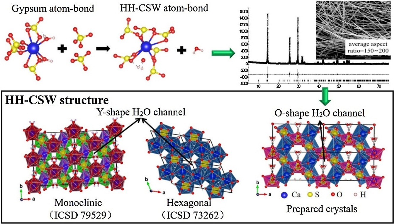
• HH-CSWs were synthesized from FGD gypsum at different holding times.
• Holding time increased sample's diffraction peaks intensity and aspect ratio.
• H2O channel's shape and size in sample are different with reported data.
• H2O released as Ca–O–H bonds broken, formed novel Ca–O–S bonds in HH-CSWs crystals.
Hemihydrate calcium sulfate whiskers (HH-CSWs) were hydrothermally synthesized in a sulfuric acid solution at 120 °C for different holding times (20, 40, and 60 min). The phase structures and morphologies were characterized by XRD and SEM, respectively. The XRD pattern of the sample under 60 min was refined via the Rietveld fitting method. The structure models of the HH-CSW sample under a 60-min holding time was established based on Rietveld fitting results. No difference in the positions of diffraction peaks was determined. The as-prepared holding time increased the intensity and aspect ratio of the diffraction peaks of both samples. In the prepared HH-CSW structure, Ca–O polyhedron is a 12-sided polyhedron similar to that in the gypsum structure; the Ca atom is located in two positions in the one-unit cell; the H2O channel along the c-axis similar is O-shaped and bigger than that in hexagonal and monoclinic CaSO4·0.5H2O structures. Therefore, although the prepared HH-CSW crystals’ structure are similar to that of the reported hexagonal and monoclinic CaSO4·0.5H2O structures, they are not the same. The formation mechanism of HH-CSW from flue gas desulfurization (FGD) gypsum is discussed based on the analysis of gypsum structure.

